
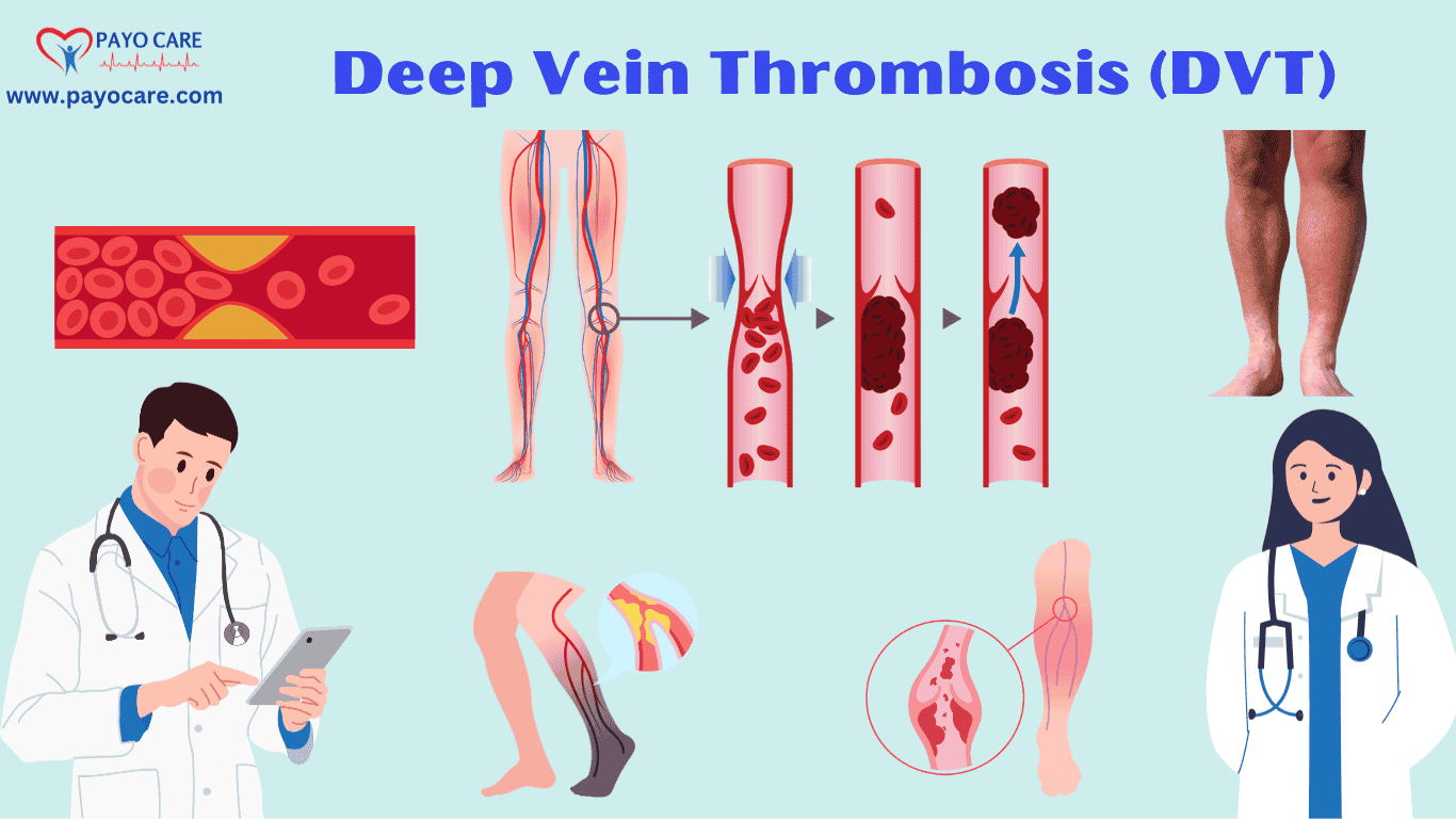

Deep Vein Thrombosis (DVT) is a serious medical condition characterized by the formation of a blood clot (thrombus) in a deep vein, typically in the legs. If left untreated, DVT can lead to life-threatening complications, such as pulmonary embolism (PE), where the clot breaks loose and travels to the lungs. Understanding the types, causes, symptoms, prevention, diagnosis, and treatments of DVT is crucial for effective management and reducing the risk of complications.
Types of DVT
1. Proximal DVT
Proximal DVT occurs in the larger, more proximal veins, such as the popliteal, femoral, or iliac veins. This type of DVT is more likely to cause significant symptoms and is associated with a higher risk of pulmonary embolism.
2. Distal DVT
Distal DVT occurs in the smaller, more distal veins, such as the calf veins. This type of DVT may cause milder symptoms and is less likely to lead to pulmonary embolism, but it can still progress to proximal DVT if not treated.
3. Upper Extremity DVT
Upper extremity DVT occurs in the deep veins of the arms, such as the subclavian, axillary, or brachial veins. This type of DVT is less common and is often associated with the use of central venous catheters or strenuous arm activity.
4. Recurrent DVT
Recurrent DVT refers to the occurrence of a new blood clot in a deep vein after a previous episode of DVT. This can happen if the underlying risk factors are not adequately managed or if anticoagulant therapy is discontinued prematurely.
Causes of DVT
DVT is caused by a combination of factors that lead to the formation of a blood clot in a deep vein. These factors are often referred to as Virchow’s triad, which includes:
1. Venous Stasis (Slow Blood Flow)
Venous stasis occurs when blood flow in the veins is slowed or stagnant. This can happen due to:
- Prolonged Immobility: Long periods of immobility, such as during long flights, bed rest, or after surgery, can lead to venous stasis.
- Heart Failure: Reduced cardiac output in heart failure can cause sluggish blood flow in the veins.
- Varicose Veins: Damaged or weakened veins can lead to poor blood flow.
2. Hypercoagulability (Increased Blood Clotting)
Hypercoagulability refers to an increased tendency of the blood to clot. This can be due to:
- Genetic Factors: Inherited conditions, such as Factor V Leiden mutation, prothrombin gene mutation, or deficiencies in natural anticoagulants (e.g., protein C, protein S, antithrombin).
- Acquired Conditions: Conditions such as cancer, pregnancy, oral contraceptive use, hormone replacement therapy, or inflammatory diseases can increase the risk of clotting.
- Surgery or Trauma: Surgery, especially orthopedic surgery, and trauma can increase the risk of blood clots.
3. Endothelial Injury (Damage to the Blood Vessel Wall)
Endothelial injury refers to damage to the inner lining of the blood vessels, which can trigger the formation of a blood clot. This can occur due to:
- Trauma: Physical injury to the veins, such as from a fracture or severe muscle injury.
- Medical Procedures: Insertion of central venous catheters or pacemakers can damage the vein walls.
- Inflammation: Inflammatory conditions, such as vasculitis, can damage the endothelium.
Symptoms of DVT
The symptoms of DVT can vary depending on the location and size of the blood clot. Common symptoms include:
1. Swelling
Swelling in the affected leg or arm is one of the most common symptoms of DVT. The swelling may be accompanied by a feeling of heaviness or tightness.
2. Pain
Pain in the affected limb is another common symptom. The pain may be described as a cramping or soreness and is often worse when standing or walking.
3. Redness and Warmth
The skin over the affected area may become red and warm to the touch due to inflammation and increased blood flow.
4. Tenderness
The affected area may be tender to the touch, and the pain may worsen with pressure.
5. Visible Veins
In some cases, the veins near the surface of the skin may become more visible or prominent.
6. Cyanosis
In severe cases, the affected limb may develop a bluish discoloration (cyanosis) due to poor blood flow.
7. Asymptomatic DVT
In some cases, DVT may be asymptomatic, especially in the early stages. This is more common with distal DVT or small clots.

Prevention of DVT
Preventing DVT involves addressing the risk factors and implementing strategies to reduce the likelihood of blood clot formation. Key prevention strategies include:
1. Lifestyle Modifications
- Regular Exercise: Regular physical activity helps to improve blood circulation and reduce the risk of venous stasis.
- Healthy Diet: A diet rich in fruits, vegetables, whole grains, and lean proteins can help to maintain a healthy weight and reduce the risk of DVT.
- Hydration: Staying well-hydrated helps to prevent blood from becoming too thick and sticky, reducing the risk of clotting.
2. Medical Interventions
- Anticoagulant Therapy: For individuals at high risk of DVT, such as those undergoing surgery or with a history of DVT, anticoagulant medications (blood thinners) may be prescribed to prevent clot formation.
- Compression Stockings: Compression stockings help to improve blood flow in the legs and reduce the risk of DVT, especially during long periods of immobility.
- Intermittent Pneumatic Compression Devices: These devices use air pressure to massage the legs and promote blood flow, reducing the risk of DVT in hospitalized patients.
3. Avoiding Prolonged Immobility
- Frequent Movement: During long flights or car rides, it is important to take breaks to stretch and move around to prevent venous stasis.
- Leg Exercises: Simple leg exercises, such as ankle rotations and calf raises, can help to promote blood flow during periods of immobility.
4. Managing Underlying Conditions
- Control of Chronic Diseases: Managing conditions such as diabetes, hypertension, and heart failure can help to reduce the risk of DVT.
- Cancer Treatment: For individuals with cancer, appropriate treatment and management of the disease can help to reduce the risk of DVT.
Diagnosis of DVT
Diagnosing DVT involves a combination of clinical assessment, imaging studies, and laboratory tests. The following diagnostic tools are commonly used:
1. Clinical Assessment
- Medical History: A detailed medical history, including risk factors, symptoms, and any previous episodes of DVT, is essential for diagnosis.
- Physical Examination: A physical examination may reveal signs such as swelling, redness, warmth, and tenderness in the affected limb.
2. Imaging Studies
- Duplex Ultrasound: Duplex ultrasound is the most commonly used imaging test for diagnosing DVT. It uses sound waves to create images of the veins and can detect the presence of a blood clot.
- Venography: Venography involves injecting a contrast dye into the veins and taking X-ray images to visualize the blood flow and detect clots. This is less commonly used due to its invasive nature.
- Magnetic Resonance Imaging (MRI): MRI can be used to detect DVT, especially in cases where ultrasound is inconclusive or when evaluating pelvic or abdominal veins.
- Computed Tomography (CT) Scan: CT scans can be used to detect DVT, particularly in the pelvis or abdomen, and to assess for pulmonary embolism.
3. Laboratory Tests
- D-Dimer Test: The D-dimer test measures a substance in the blood that is released when a blood clot breaks down. A positive D-dimer test indicates the presence of a clot, but it is not specific to DVT and can be elevated in other conditions.
- Coagulation Studies: Blood tests to assess clotting factors, such as prothrombin time (PT) and activated partial thromboplastin time (aPTT), may be performed to evaluate the risk of clotting.

Treatments for DVT
The primary goal of DVT treatment is to prevent the clot from growing, reduce the risk of pulmonary embolism, and prevent recurrence. Treatment options include:
1. Anticoagulant Therapy
Anticoagulant medications, also known as blood thinners, are the mainstay of DVT treatment. They work by preventing the formation of new clots and allowing the body to dissolve the existing clot. Common anticoagulants include:
- Heparin: Heparin is often used as an initial treatment for DVT, either as an intravenous infusion or subcutaneous injection. Low molecular weight heparin (LMWH), such as enoxaparin, is commonly used due to its ease of administration.
- Warfarin: Warfarin is an oral anticoagulant that is often used for long-term treatment of DVT. It requires regular monitoring of the International Normalized Ratio (INR) to ensure the correct dosage.
- Direct Oral Anticoagulants (DOACs): DOACs, such as rivaroxaban, apixaban, dabigatran, and edoxaban, are newer oral anticoagulants that do not require regular monitoring and have fewer dietary restrictions compared to warfarin.
2. Thrombolytic Therapy
Thrombolytic therapy, also known as clot-busting therapy, involves the use of medications to dissolve the blood clot. This is typically reserved for severe cases of DVT, such as those causing significant limb ischemia or massive pulmonary embolism. Thrombolytics, such as alteplase, are administered intravenously or directly into the clot via a catheter.
3. Inferior Vena Cava (IVC) Filter
In cases where anticoagulant therapy is contraindicated or ineffective, an IVC filter may be placed in the inferior vena cava to prevent clots from traveling to the lungs. The filter is typically inserted through a minimally invasive procedure and can be temporary or permanent.
4. Compression Therapy
Compression stockings or bandages are often used to reduce swelling and improve blood flow in the affected limb. Compression therapy can also help to prevent post-thrombotic syndrome, a complication of DVT characterized by chronic pain and swelling.
5. Surgical Thrombectomy
Surgical thrombectomy involves the removal of the blood clot through a surgical procedure. This is typically reserved for severe cases of DVT, such as those causing significant limb ischemia or when thrombolytic therapy is contraindicated.
6. Lifestyle and Long-Term Management
- Regular Follow-Up: Regular follow-up with a healthcare provider is essential to monitor the effectiveness of treatment and adjust medications as needed.
- Lifestyle Changes: Adopting a healthy lifestyle, including regular exercise, a balanced diet, and avoiding prolonged immobility, can help to reduce the risk of recurrent DVT.
- Management of Underlying Conditions: Managing underlying conditions, such as cancer, heart failure, or inflammatory diseases, is important to reduce the risk of recurrent DVT.
Conclusion
Deep Vein Thrombosis (DVT) is a serious condition that requires prompt diagnosis and treatment to prevent life-threatening complications such as pulmonary embolism. Understanding the types, causes, symptoms, prevention, diagnosis, and treatments of DVT is crucial for effective management and reducing the risk of complications. By addressing risk factors, implementing preventive measures, and adhering to appropriate treatment plans, individuals with DVT can achieve better outcomes and improve their quality of life.
If you suspect that you or someone else may have DVT, it is important to seek medical attention immediately. Early diagnosis and treatment are key to preventing complications and ensuring a successful recovery.
Disclaimer: This article is for informational purposes only